
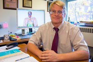
A team of electrical and computer engineers at Worcester Polytechnic Institute (WPI) has created a virtual human by digitizing and combining high-resolution images of thin slices of an actual human body.
The highly detailed digital model, known as a computational human phantom, is a tool that can be used to conduct virtual medical procedures and medical experiments—including procedures too risky to perform on living people—without the need for actual human subjects. The model will fill a specific need, and its availability is anticipated by the medical community. To ensure the technology’s effectiveness for those purposes, WPI researchers have teamed up with faculty at Harvard Medical School.
Developed by Sergey Makarov, professor of electrical and computer engineering at WPI, along with research scientist Gregory Noetscher ’00, ’14 (PhD); PhD candidate Janakinadh Yanamadala, and several undergraduates majoring in electrical engineering and computer science, the model is the product of more than four years of painstaking work using proprietary software developed under Makarov’s guidance.
The model was developed by combining high-resolution color photographs of 5,000 cross-sectional slices (each a third of a millimeter thick) of a human cadaver—a woman from Maryland who had donated her body to science. The images were created for the National Library of Medicine, which made them available to Makarov. He and his team used a variety of image processing techniques to align the images and digitally stitch them into a highly detailed three-dimensional virtual human body.
The virtual body was then transformed into a finite element model that contains detailed information about the location and characteristics of the many tissues that make up the body’s organs and systems. This model can be used as a computational tool to study how the body will respond to specific treatments and medical procedures.
"There are many cases where doctors need to investigate what is happening in the human body, but it would be too difficult to do the corresponding experiments," Makarov said. "This process would effectively help doctors and other medical researchers."

For example, collaborating with Ara Nazarian, assistant professor of orthopaedic surgery at Harvard Medical School and Beth Israel Deaconess Medical Center’s Center for Advanced Orthopaedic Studies, Makarov and his team have used the model to explore how tissues surrounding metal implants, including replacement joints and rods or nails used to repair fractures, will respond if a patient is placed in an MRI scanner.
It might be dangerous, Makarov noted, for an elderly person with a metal hip implant to undergo an MRI scan since that metal will be heated by the combination of a strong magnetic field and radio waves used to create MRI images. By incorporating a model of the implant within the virtual human, the researchers can test the body’s response to the heating during various imaging protocols without exposing a human volunteer to any risk. "This would allow doctors to perform any number of tests safely and without hurting humans," said Makarov.
The virtual human model has been optimized to be compatible with software that computes electromagnetic activity for analysis in a variety of applications. Such applications could ultimately lead to better medical procedures for humans and updated guidance on radiation exposure. For example, Makarov and his students are using the model to evaluate the effectiveness of electrodes that can treat Parkinson’s disease with electrical stimulation and exploring various configurations of an antenna that may be used to treat tumors with electromagnetic radiation.
"This approach allows you to model things beforehand, whether that means exploring how a body would react in an MRI machine or how certain diseases may react differently to a device or diagnostic modality," said Nazarian, whose role in the research is to oversee how the human model might be used in clinical applications. "The model lets you see how close you can explore various studies in a simulated medical environment."
Nazarian noted that such virtual experiments are much easier to conduct and are far less costly than actual clinical trials. In addition, he said, the computational human models may help improve patient care if it can be used to help diagnose diseases and evaluate treatment plans.
While much of the work being conducted with the model is virtual, the model can take physical form. In fact, in one process, Makarov used the finite element model of the virtual human’s femur to create a CAD model that he could output, to scale, on a 3D printer. Such physical representations of body parts might also be useful in medical and other physiological research.
Makarov and Nazarian are currently seeking to license the virtual human software and make it available to research universities and other medical groups. "Professor Makarov’s team and I have collaborated very effectively on this project and we are excited to see this work come to fruition," said Nazarian.
The model development was completed by WPI’s Electrical and Computer Engineering Department and NEVA Electromagnetics, LLC, a private company in Yarmouth Port, Mass., founded by Makarov. Additional support was provided by the Department of Neurophysics at Max Planck Institute for Human Cognitive and Brain Sciences in Germany.


