Three neuroscience researchers investigating Alzheimer's disease are pictured over a logo for Worcester Polytechnic Institute.

From left, Senbao Lu, Bhaavin Jogeshwar, and Benjamin Nephew

Predicting Alzheimer’s Disease

WPI researchers use machine learning and brain scans to identify age- and sex-specific anatomical patterns that predict disease
March 5, 2026

WPI researchers have used a form of artificial intelligence (AI) to analyze anatomical changes in the brain and predict Alzheimer’s disease with nearly 93% accuracy. 

Preview

Computerized images of brains are displayed on a grid.

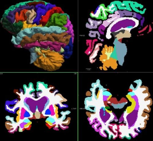
Segmented and labeled images of a normal brain

Their research, published in the journal Neuroscience, also revealed that the anatomical changes, involving loss of brain volume, differ by age and sex. 

“Early diagnosis of Alzheimer’s disease can be difficult because symptoms can be mistaken for normal aging,” says Benjamin Nephew, assistant research professor in the Department of Biology and Biotechnology. “We found that machine-learning technologies, however, can analyze large amounts of data from scans to identify subtle changes and accurately predict Alzheimer’s disease and related cognitive states. This advance has informed Alzheimer’s disease research and may lead to methods that could allow doctors to diagnose and treat the disease earlier and more effectively.”

Alzheimer’s disease is a neurodegenerative disorder that impairs mental functions and ultimately leads to death. An estimated 6.9 million Americans age 65 and older are living with Alzheimer’s disease.

Healthy brains contain billions of neurons, the cells that process and transmit signals needed for thought, movement, and other bodily functions. Alzheimer’s disease injures neurons, leading to cell death and loss of brain tissue and associated cognitive functions.

Nephew, PhD student Senbao Lu, and Bhaavin Jogeshwar, MS ’24, conducted their research with MRI scans of brains from the Alzheimer’s Disease Neuroimaging Initiative, a multicenter project that built a library of brain scans from people age 69 to 84. The scans depict the brains of people with normal mental functioning, mild cognitive impairment, and Alzheimer’s disease.

Analyzing data-rich MRI images can require substantial computing power and time. To focus their investigation, the WPI researchers first used machine learning to analyze 815 MRI scans for volume measurements in 95 brain regions. Then they deployed an algorithm to make predictions based upon differences in the measurements between healthy individuals and those with mild cognitive impairment or Alzheimer’s disease.

Results showed that the method was 92.87% accurate in detecting Alzheimer’s disease among normal brains and brains of people with mild cognitive impairment. 

Beginning Quote Icon of beginning quote
The critical challenge in this research is to build a generalizable machine-learning model that captures the difference between healthy brains and brains from people with mild cognitive impairment or Alzheimer’s disease. Beginning Quote Icon of beginning quote
  • Benjamin Nephew
  • Assistant Research Professor, Department of Biology and Biotechnology

Volume loss in the hippocampus, amygdala, and entorhinal cortex were top predictors of Alzheimer’s disease across age and sex categories. The hippocampus is a small seahorse-shaped structure deep in the brain that is responsible for memory and learning. The amygdala, which is made up of two almond-shaped structures, controls emotions. The entorhinal cortex is a hub for memory, navigation, and perception, and it is among the first parts of the brain to be impacted by Alzheimer’s disease.

Both males and females age 69 to 76, the youngest age group studied, showed loss of brain volume in the right hippocampus. The researchers say that suggests the right hippocampus may be important in early diagnosis of Alzheimer’s disease.

“The critical challenge in this research is to build a generalizable machine-learning model that captures the difference between healthy brains and brains from people with mild cognitive impairment or Alzheimer’s disease,” Nephew says. “A generalizable model means that the biomarkers we found are not unique to this dataset but could be universal to all patients with mild cognitive impairment or Alzheimer’s.”

Differences in male and female brains also emerged. The researchers discovered that volume loss in females occurred in the left middle temporal cortex, which is involved in language, memory, and visual perception. In males, volume loss was notable in the right entorhinal cortex.

The degree of these differences was surprising, Nephew says, and may be related to interactions between the progression of Alzheimer’s disease and changes in sex hormones. Some researchers have connected the risk of Alzheimer’s disease to the loss of estrogen in women and testosterone in men as they age.

Nephew and WPI students are following up on their Neuroscience publication by evaluating the use of deep leaning models and examining other factors that may impact the brain and Alzheimer’s disease, such as diabetes. The research has attracted WPI students from disciplines ranging from biology and biotechnology to neuroscience, psychology, computer science, and bioinformatics.

“This research exemplifies the strength of neuroscience at WPI, which is interdisciplinary and computational,” Nephew says. “The brain is an extremely complicated organ, and we need to think broadly about how to better understand, predict, and treat the diseases that afflict the brain.”

Keep Up With WPI Research News

Want to learn more about Research at WPI? Subscribe to the monthly newsletter.


Explore WPI's MS in Neuroscience

Profiles