bio

Robotic System Aims for Better Cancer Detection

A novel robotic system promises to make prostate cancer biopsies faster, more accurate, less costly, and less discomforting for the patient.
August 24, 2016

A novel robotic system may help transform the detection of prostate cancer, the last form of cancer still diagnosed with blind needle biopsies. The heart of the system is a specially designed MRI-compatible robot that can work inside the bore of an MRI scanner to help a physician guide a biopsy needle precisely toward a target of interest identified using real-time images. It promises to make prostate cancer biopsies faster, more accurate, less costly, and less discomforting for the patient.

The robotic system was developed by a team of WPI robotics engineers led by Gregory Fischer, PhD, associate professor of mechanical engineering and robotics engineering and director of WPI’s Automation and Interventional Medicine (AIM) Laboratory. The work has been carried out in collaboration with researchers at Johns Hopkins University, Brigham and Women’s Hospital in Boston, and Acoustic MedSystems Inc.

The system is currently being tested as part of a larger clinical research program at Brigham and Women’s funded by a Bioengineering Research Partnership award from the National Institutes of Health through the National Cancer Institute. The program aims to replace blind needle biopsies with image-guided technology, notes Clare Tempany, MD, professor of radiology at Harvard Medical School, chair of research radiology at Brigham and Women’s, and principal investigator for the research program. “The ultimate goal of our group is to develop enabling technologies that extend the capabilities of physicians to treat their patients.”

Most prostate biopsies today are performed with the aid of ultrasound, which can localize the prostate but not readily detect potential cancers. Requiring multiple needle insertions, the biopsies suffer from low sensitivity and often misleading results. In fact, about 35 percent of serious tumors may be missed during initial biopsies.

PROFILE(S)
This technology should permit greater accuracy, and the odds of hitting the target on the first try should be higher.
  • Gregory Fischer
  • Associate Professor of Mechanical Engineering and Robotics Engineering
  • Director, WPI’s Automation and Interventional Medicine (AIM) Laboratory

In the Brigham and Women’s program, biopsies are being performed with the aid of real-time MRI imaging — both with and without the MRI-compatible robot. When working without the robot, physicians use a plastic grid to help position the biopsy needle. They first use multimodality MRI scans to generate a plan showing where the needle should be inserted. Then, with the patient in the MRI scanner, the physician directs the needle through most appropriate guide holes in the grid. Additional scans are made periodically to verify the path of the needle and make adjustments, if needed.

Rather than restrict the needle positioning to the choices offered by a grid, the robot manipulates a needle-guide inside the bore of the scanner to help the physician place the needle in the most optimal position as indicated by the real-time images generated by the MRI. “The robot gives the physician a great deal more choice about where to place the biopsy needle,” Fischer says. “This technology should permit greater accuracy, and the odds of hitting the target on the first try should be higher.”

This “first-in- human” testing of the robotic system is the culmination of more than six years of research and development by Fischer, who, along with Julian Lordachita and colleagues in the Laboratory for Computational Sensing and Robotics (LCSR) at Johns Hopkins University, has pioneered the development of compact, high precision surgical robots that are expressly designed to work in the environment inside the bore of an MRI scanner, as well as the electronic control systems and software needed to operate the robots with the safety, reliability, and ease of use required of technology designed for the operating room.

The ultimate goal of our group is to develop enabling technologies that extend the capabilities of physicians to treat their patients.
  • Clare Tempany, MD
  • Professor of Radiology, Harvard Medical School
  • Chair of Research Radiology at Brigham and Women’s


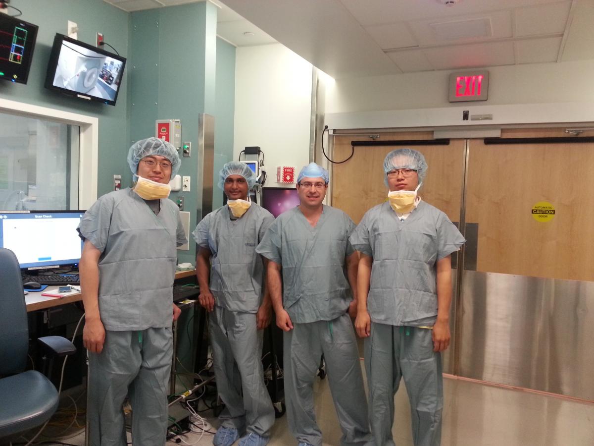
Gregory Fischer, second from right, with PhD candidates, from left,

Weijian Shang, Nirav Patel, and Gang Li, in the Advanced Multimodality

Image-Guided Operating suite at Brigham and Woman’s Hospital

To develop robots that can work inside an MRI scanner, Fischer and his team have had to overcome several significant technical challenges. Most important, since the scanner includes a powerful magnet, the robot, including all of its sensors and actuators, must be made from nonferrous materials. The robot used in the prostate cancer trial is built primarily of plastic parts and uses ceramic piezoelectric motors.

This article originally appeared in the 2015 edition of WPI Research.